A Stress-Induced Vicious Cycle in the Development of T1D
Contact PI: Peter Arvan, PhD, University of Michigan (U01 DK127747)
Leslie Satin, PhD, Investigator, University of Michigan
Scott Soleimanpour, PhD, Investigator, University of Michigan
Start Date: September 15, 2020
End Date: December 2025
Abstract
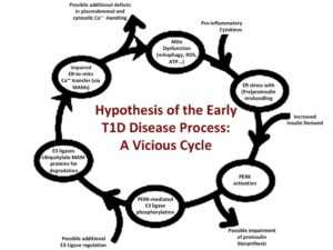
A vicious cycle of (pre)proinsulin-mediated ER stress, ubiquitin-ligase activation, and disordered Ca2+- handling in pancreatic beta cells. This proposal is submitted in response to a request-for-applications for Discovery of Early Type 1 Diabetes Disease Processes in the Human Pancreas (including the possibility of studies of signaling/processing pathways that are dysregulated in stressed beta cells during the asymptomatic phase of T1D). Our group of three pancreatic beta cell biologists with distinct expertise (Drs. Arvan, Soleimanpour, and Satin) brings forward a novel hypothesis about the initiating beta cell events in T1D, with the realization that these intricacies exceed what any one laboratory could exhaustively study on their own. Specifically, the three collaborating P.I.s propose a chain of beta cell defects that can be initiated and exacerbated by pro- inflammatory stimuli but are then further propagated by a failure of inter-organellar support, including the endoplasmic reticulum (ER) and mitochondria. It is well known that activation of ER stress signaling upon perturbation of ER homeostasis (launched from multiple potential initiators, including proinflammatory cytokines) triggers activation of stress kinases that include (but are not limited to) the ER membrane protein, PERK. New evidence suggests that ER stress kinase activity results in phosphorylation (and thereby activation) of one or more E3 ubiquitin (Ub) ligases residing at ER-mitochondrial contact sites known as MAMs. We posit that ER stress-related E3 Ub-ligase activation stimulates ubiquitylation and proteasomal turnover of a number of MAM resident proteins including the well-recognized mitochondrial substrate, mitofusin-2, but also key components of ER-to-mitochondrial Ca2+ transfer including IP3R1 and others. ER stress-provoked degradation of MAM protein components can be evaluated biochemically and quantitatively by Western blotting; but we go beyond this to examine the functional consequences via measurements of impaired ER-to- mitochondrial Ca2+ transfer. We propose that such calcium imbalance further impairs mitochondrial function including (but not limited to) impaired ATP generation, which exacerbates the perturbation of ER homeostasis with concomitant ER stress. In short, we propose that pro-inflammatory triggers of T1D can initiate a vicious cycle leading to beta cell failure. This hypothesis will be tested in human islets in vitro as well as in an in vivo model involving transplanted human islets. Finally, we propose to probe certain trigger points to break the vicious cycle, in an attempt to rescue beta cell survival and function, with the intention of preventing T1D onset and progression.

Publications
- TRAF6 integrates innate immune signals to regulate glucose homeostasis via Parkin-dependent and Parkin-independent mitophagy
- Exendin-4 enhances insulin-positive phenotype of human pluripotent stem cell-derived β cells during transplantation
- TRAF6 integrates innate immune signals to regulate glucose homeostasis via Parkin-dependent and -independent mitophagy
- Retrograde mitochondrial signaling governs the identity and maturity of metabolic tissues
- Role of Sec61α2 translocon in insulin biosynthesis
- Limitations in mitochondrial programming restrain the differentiation and maturation of human stem cell-derived β cells
- A metabolic redox relay supports ER proinsulin export in pancreatic islet β-cells
- Untangling the genetics of beta cell dysfunction and death in type 1 diabetes
- LONP1 regulation of mitochondrial protein folding provides insight into beta cell failure in type 2 diabetes
- Do We Need a New Hypothesis for KATP Closure in β-Cells? Distinguishing the Baby From the Bathwater
- Proinsulin folding and trafficking defects trigger a common pathological disturbance of endoplasmic reticulum homeostasis
- Autophagy of the ER: the secretome finds the lysosome
- A versatile pumpless multi-channel fluidics system for maintenance and real-time functional assessment of tissue and cells
- K(ATP) channel activity and slow oscillations in pancreatic beta cells are regulated by mitochondrial ATP production
- Deconstructing the integrated oscillator model for pancreatic β-cells
- Complementary Approaches to Interrogate Mitophagy Flux in Pancreatic β-Cells
- SERCA2 regulates proinsulin processing and processing enzyme maturation in pancreatic beta cells
- Protein disorder in the regulatory control of mitophagy
- Mitochondrial metabolism and dynamics in pancreatic beta cell glucose sensing
- Extrahepatic transplantation of 3D cultured stem cell-derived islet organoids on microporous scaffolds
- Ca(2+) Release or Ca(2+) Entry, that is the Question: What Governs Ca(2+) Oscillations in Pancreatic Beta Cells?
- Simultaneous Measurement of Changes in Mitochondrial and Endoplasmic Reticulum Free Calcium in Pancreatic Beta Cells
- Reciprocal regulatory balance within the CLEC16A-RNF41 mitophagy complex depends on an intrinsically disordered protein region
- Evaluation of polygenic risk scores to differentiate between type 1 and type 2 diabetes
- Editorial: Study of pancreatic islets based on human models to understand pathogenesis of diabetes
- β-Cell Cre Expression and Reduced Ins1 Gene Dosage Protect Mice From Type 1 Diabetes
- Effects of Pro-Inflammatory Cytokines on the Pancreatic Islet Transcriptome: A View at Both Temporal and Single-Cell Resolution
- Carbonyl Posttranslational Modification Associated With Early-Onset Type 1 Diabetes Autoimmunity
- Adaptation to chronic ER stress enforces pancreatic β-cell plasticity
- Mitofusins Mfn1 and Mfn2 Are Required to Preserve Glucose- but Not Incretin-Stimulated β-Cell Connectivity and Insulin Secretion
- An intrinsically disordered protein region encoded by the human disease gene CLEC16A regulates mitophagy
- Mitofusin 1 and 2 regulation of mitochondrial DNA content is a critical determinant of glucose homeostasis
- Pulsatile Basal Insulin Secretion is Driven by Glycolytic Oscillations
- Beta-Cell Ion Channels and Their Role in Regulating Insulin Secretion
- New Aspects of Diabetes Research and Therapeutic Development
- Roles of Calreticulin in Protein Folding, Immunity, Calcium Signaling and Cell Transformation
- A Selective Look at Autophagy in Pancreatic β-Cells
- Cell Cycle Regulation of the Pdx1 Transcription Factor in Developing Pancreas and Insulin-Producing β-Cells
- Mitophagy protects beta cells from inflammatory damage in diabetes

