Spatial Profiling of Pancreatic N-Glycosylation in T1D
Contact PI: Jordan Wright, MD, PhD, Vanderbilt University (R03 147555)
Start Date: May 1, 2026
NIH HIRN Gateway Investigator Award Recipient
Abstract
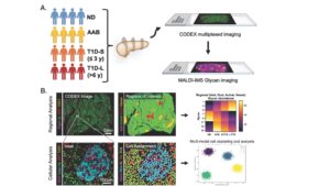
Type 1 diabetes (T1D) is caused by T-cell mediated destruction of insulin-producing pancreatic beta cells. While notable progress has been made in predicting and delaying onset of T1D, our limited understanding of the factors that initiate and maintain this autoimmune attack continue to act as a major barrier to further progress. Increasing evidence points to the pancreas itself, including endocrine cells, exocrine cells, and the extracellular matrix, as possible contributors to the pathogenic immune activation. One factor that is known to contribute to immune cell activation, and that is altered in other pancreatic disease including cancer, is protein N-glycosylation, wherein complex carbohydrate chains called glycans are enzymatically attached to specific asparagine (called N-glycans) residues as proteins transit the endoplasmic reticulum and golgi complex. Glycosylation patterns influence protein stability, localization, and receptor binding, which can dramatically alter cell function and intercellular communication. Though the pancreatic glycome has been studied in pancreatic cancer, very little is known about how the glycome changes in diabetes pathogenesis, partially owing to the relative scarcity of appropriate human tissues to study and to the complexity of analysis methods required to measure protein glycosylation. While single cell transcriptomic data shows that expression of many of the enzymes involved in glycosylation are altered in T1D, it remains unknown how the pancreatic glycome changes during T1D pathogenesis, and whether altered glycosylation contributes to changes in pancreatic structure, cell composition, or immune cell infiltration. I hypothesize that N-glycosylation in the pancreas is altered as type 1 diabetes progresses, contributing to changes in immune cell localization and phenotype. I will employ two state-of-the-art imaging technologies to test this hypothesis in pancreas tissues from donors without diabetes, with positive auto-antibodies, or with recent-onset or long-standing T1D: 1) Imaging mass spectrometry will allow comprehensive quantitation of different N-glycans across entire tissue sections and at single-cell resolution, and 2) Multiplex immunofluorescence microscopy (CODEX) will be used to define pancreatic regions of interest and to quantify cell types and subtypes across the same tissue section. In Aim 1, I will test the hypothesis that the pancreatic N-glycome quantitatively changes throughout T1D progression. In Aim 2, I will test the hypothesis that regions of altered N-glycome signature are associated with changes in cellular composition and immune cell phenotypes. Completion of these aims will identify high level changes to post-translational protein processing signatures as T1D progresses. These results will lay the groundwork for future studies into mechanisms responsible for glycomic changes, identification of specific proteins that are affected, and definition of novel glycoprotein signatures that may be promising biomarkers or drug targets.
 Follow


